What does GMS Stain detect
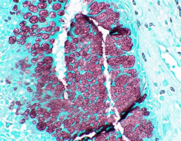
Grocott methenamine silver (GMS) stain is commonly used for the identification of fungi on cytosmears and tissue sections. It imparts a black color to the fungal profiles and a pale green color to the background. It stains all pathogenic and nonpathogenic fungi and melanin.
Do bacteria stain with GMS?
GMS is known to stain bacteria and has been shown to be useful in identifying bacteria in prosthetic valve endocarditis. This example is presented to emphasise the utility of Grocott methenamine silver staining in the identification of bacteria.
What is silver staining used for?
Silver staining is the most sensitive colorimetric method for detecting total protein. The technique involves the deposition of metallic silver onto the surface of a gel at the locations of protein bands. Silver ions (from silver nitrate in the staining reagent) interact and bind with certain protein functional groups.
What is gomori methenamine stain used for?
Gomori’s Methenamine Silver (GMS) stain is used for fungi and bacteria. The fungi and bacteria are turned black, while everything else is stained green with Light green SF solution.What does Ziehl Neelsen stain?
Ziehl–Neelsen staining is a bacteriological stain used to identify acid-fast organisms, mainly Mycobacteria. It is named for two German doctors who modified the stain: the bacteriologist Franz Ziehl (1859–1926) and the pathologist Friedrich Neelsen (1854–1898).
Which stain is used for fungus?
The Grocott’s silver (GMS) stain is probably the most widely used fungal stain, however, the somewhat capricious nature of silver staining may challenge even the most experienced of histology practitioners.
How does acid-fast stain work?
Some of the sample is placed on a glass slide, stained, and heated. The cells in the sample hold onto the dye. The slide is then washed with an acid solution and a different stain is applied. Bacteria that hold onto the first dye are considered “acid-fast” because they resist the acid wash.
Which stain is used for AFB staining?
The acid-fast bacilli will stain bright red, and the background will stain blue. Reagents used in the procedure include Ziehl–Neelsen carbol-fuchsin solution, 1% acid alcohol, and methylene blue solution [15].What is Gram staining an example of?
Gram staining is a bacteriological laboratory technique used to differentiate bacterial species into two large groups (gram-positive and gram-negative) based on the physical properties of their cell walls.
Why is dried smear stained with methylene blue?It is a cationic dye that stains cells blue because the positively charged dye is attracted to negatively charged particles such as polyphosphates, DNAs, and RNAs. Specimens collected from patients by swabbing are smeared onto microscope slides and the methylene blue solution is dropped on the slide.
Article first time published onHow do you make methenamine silver?
- Hydrate sections with distilled water.
- Then oxidize the section with 4% aqueous chromic acid at room temperature and leave for 1 hr.
- Wash in water for a few seconds.
- Treat the sections with 1% sodium metabisulphite for 1 min.
- Wash in smoothly running tap water for 3 mins.
Which Colour is obtained after PAS in glycogen containing tissue?
However, detection of glycogen in cells and tissues has long been achieved by histochemical methods, the most common of which is PAS staining whereby oxidation of hydroxyls in glucose residues by periodic acid is followed by reaction of the resulting aldehydes with Schiff reagent to generate a purple or magenta color ( …
What tissue does silver stain stain?
Identifies reticulin fibers in tissue sections liver, kidney and spleen. Reticulin is a type III collagen found in the basement membrane of many organs and provides structural integrity. Silver staining is a highly sensitive method for detecting proteins in polyacrylamide slab gels.
Is silver staining more sensitive than Coomassie?
Coomassie Blue staining is approximately 50-fold less sensitive than silver staining, however due to its simplicity binding Coomassie Blue is preferred. … Staining methods that allow destaining of gels makes them more appealing, as destaining may be needed for certain downstream applications.
What stains myelin?
The first method is conventional luxol fast blue (LFB) method which stains myelin in blue and Nissl bodies and mast cells in purple.
How do you use Ziehl-neelsen stain?
- Spread the sputum evenly over the central area of the slide using a continuous rotational movement. …
- Place slides on the dryer with smeared surface upwards, and air dry for about 30 minutes.
- Heat fix dried smear.
- Cover the smear will carbol fuchsin stain.
What is the difference between Gram staining and Ziehl-Neelsen staining?
An acid-fast stain is able to differentiate two types of gram-positive cells: those that have waxy mycolic acids in their cell walls, and those that do not. Two different methods for acid-fast staining are the Ziehl-Neelsen technique and the Kinyoun technique. Both use carbolfuchsin as the primary stain.
Is Gram stain an adequate substitute for acid fast stain Why?
Is a gram stain an adequate substitute for an acid fast stain? … No, it won’t hold the stain due to the mycolic acid lipid.
Why is acid fast staining not used as widely as Gram staining?
Why is the acid fast stain not as widely used as the gram stain? … very few bacteria are acid fast positive, so the test is less useful than a gram stain, which separates organisms into two large groups.
Why is a mordant used in the Gram stain?
The function of a mordant in a Gram stain is to prevent the crystal violet from leaving the Gram-positive cell.
Can fungi be Gram stained?
Fungi (in the form of yeasts or molds) may be seen on a Gram stain and are reported. Yeast may appear as single cells that may have buds, while molds may appear as a wide variety of plant-like branches called hyphae. Further testing may be performed to identify the specific type.
What is staining Slideshare?
STAIN: Stain is a dye used to color the living or dead organelles. 3. TYPES: ACIDIC: Negatively charged acid radicals imparts color in eosin, acid fuchsine, malachite green, nigrosin, Indian ink. BASIC: Positively charged basic radicals combines with negatively charged particles in cytoplasm and gives color.
Is blastomycosis Gram positive or negative?
General fungal culture returned negative after 72 h incubation. However, NGS of both lavage and biopsy specimen revealed the presence of Blastomyces dermatitidis. Gram stain of the lavage specimen showed a gram-positive broad-based budding yeast, and immunofluorescence also revealed Blastomyces dermatitidis (Fig. 3).
What is the importance of Gram staining in microbiology?
The Gram stain is the most important staining procedure in microbiology. It is used to differentiate between gram positive organisms and gram negative organisms. Hence, it is a differential stain. Gram negative and gram positive organisms are distinguished from each other by differences in their cell walls.
Is Gram staining a simple stain?
The Gram stain is a differential stain, as opposed to the simple stain which uses 1 dye. As a result of the use of 2 dyes, making this procedure a differential stain, bacteria will either become purple/blue or pink during the procedure.
What is difference between gram positive and negative?
Gram positive bacteria have a thick peptidoglycan layer and no outer lipid membrane whilst Gram negative bacteria have a thin peptidoglycan layer and have an outer lipid membrane.
What are the steps of Gram staining?
The performance of the Gram Stain on any sample requires four basic steps that include applying a primary stain (crystal violet) to a heat-fixed smear, followed by the addition of a mordant (Gram’s Iodine), rapid decolorization with alcohol, acetone, or a mixture of alcohol and acetone and lastly, counterstaining with …
Is methylene blue used in Gram staining?
Often the first test performed, gram staining involves the use of crystal violet or methylene blue as the primary color. … Initially, all bacteria take up crystal violet dye; however, with the use of solvent, the lipid layer from gram-negative organisms is dissolved.
What organelles does methylene blue stain?
Methylene blue stains negatively charged molecules in the cell, including DNA and RNA. This dye is toxic when ingested and it causes irritation when in contact with the skin and eyes. The cells seen are squamous epithelial cells from the outer epithelial layer of the mouth.
Is Gram stain romanowsky?
Romanowsky-type stains, new methylene blue, and Gram stain are all satisfactory for making a cytologic diagnosis.
How do you make stained GMS?
- Bring sections to distilled water.
- Place slides in 40 ml 4% aq chromic acid in a loosely covered plastic coplin jar. …
- Dip slides up and down in coplin jar and leave to stand for a further 2 min.
- Wash in running tap water 30 sec.
- Treat sections with 1% sodium metabisulphite 30 sec.