What does Schistocytes mean
Schistocytes or schizocytes are defined as circulating red blood cell fragments. Detection of schistocytes is an important clue for the diagnosis of thrombotic microangiopathy (TMA), which includes both thrombotic thrombocytopenic purpura (TTP) and hemolytic-uremic syndrome (HUS).
What is the significance of schistocytes?
Schistocytes or schizocytes are defined as circulating red blood cell fragments. Detection of schistocytes is an important clue for the diagnosis of thrombotic microangiopathy (TMA), which includes both thrombotic thrombocytopenic purpura (TTP) and hemolytic-uremic syndrome (HUS).
Where are schistocytes found?
Schistocytes, or “split” red cells, appear in the peripheral blood smear (>1% of total red cells)2 as an indication of the microangiopathic hemolytic anemia.
What causes fragmented red blood cells?
The most common causes of abnormal fragmentation were malignancy with cytotoxic chemotherapy and severe iron deficiency. In two subjects, an abnormal red blood cell fragmentation pattern was the clue to a spectrin mutant in subjects with an automated blood count previously evaluated as normal.What does 1+ schistocytes mean?
The presence of ≥1% schistocytes on a peripheral blood smear (PBS) is an important criterion for the diagnosis of thrombotic microangiopathy (TMA). The reporting of schistocytes has been standardized by the International Council for Standardization in Hematology (ICSH).
What causes schistocytes in TTP?
Thrombotic thrombocytopenic purpura Platelets end up being removed and the resulting fibrin strand formation remains. These fibrin strands along with the stress from the blood flow cause fragmentation of the red blood cells, leading to schistocyte formation.
Are schistocytes the same as helmet cells?
Some of the irregular shapes appear as “helmet” cells. Such fragmented RBC’s are known as “schistocytes” and they are indicative of a microangiopathic hemolytic anemia (MAHA) or other cause for intravascular hemolysis.
How do I report schistocytes?
Schistocytes shall be counted on PB smears using an OM at medium (x400) or high (x1000) magnification and expressed as a percentage of RBCs after counting at least 1,000 RBCs (confirmed).When do you see schistocytes?
Schistocytes are likely to be seen in hemolytic anemias, especially microangiopathic hemolytic anemia in which there is mechanical trauma to erythrocytes attempting to pass through fibrin strands in small vessels. Patients usually also have thrombocytopenia.
Are schistocytes present in DIC?Discussion: Schistocytes were thus frequently observed in DIC patients, usually with low percentage, within or close to the reference range (<0.5%).
Article first time published onWhat are Acanthocytes?
Acanthocytosis is a red cell phenotype associated with various underlying conditions. Acanthocytes (from the Greek word acantha, which means thorn), or spur cells, are spiculated red cells with a few projections of varying size and surface distribution (see the images below).
Why is PNH nocturnal?
For some time, paroxysmal nocturnal hemoglobinuria (PNH) has been known to result from somatic mutations in the PIGA gene, which encodes phosphatidylinositol glycan class A (PIGA). These mutations result in hematopoietic stem cells that are deficient in glycosyl-phosphatidylinositol anchor protein (GPI-AP).
Are schistocytes seen in PNH?
Results: Anemia and/or leukopenia and/or thrombocytopenia, increased reticulocyte count and LDH were observed in patients with PNH clone. Some of them had dacriocytes, schistocytes.
Do schistocytes have central pallor?
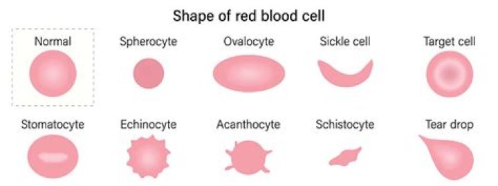
Schistocytes are red cell fragments that may be present in microangiopathic hemolysis and other causes of mechanical hemolysis. Schistocytes are smaller than red blood cells, lack central pallor, and have sharp angles and/or straight borders. … Spherocytes are also frequently seen when schistocytes are present.
Can you have DIC without schistocytes?
The presence of schistocytes is neither sensitive nor specific for DIC, but in certain instances, it may help confirm a chronic DIC diagnosis when the schistocytes are seen in concert with normal coagulation values and increased D-dimer levels.
Can dialysis cause Schistocytes?
We have observed many patients with no other identifiable cause for schistocyte formation who are receiving dialysis. Schistocytes are fragmented red cells which may take a variety of shapes and are usually associated with mechanical disruption of the red cell membrane.
How many Schistocytes are significant per HPF?
Two (2) schistocytes per HPF correlated with 1% schistocytes on the linear plot. At UCMC, policy had been to report 2-8 schistocytes per HPF as present and >8 per HPF as increased. These findings indicated that the threshold for reporting increased schistocytes should be lowered from >8 per HPF to >2 per HPF.
Does sickle cell cause schistocytes?
Described is a case of acute chest syndrome in a sickle- cell patient (hemoglobin SS) who also developed signs and symptoms of thrombotic thrombocytopenic purpura, including thrombocytopenia and hemolysis (anemia, elevated lactate dehydrogenase, presence of schistocytes, dark-colored plasma, and elevations in nucleated …
What causes nucleated red blood cells in adults?
The presence of nucleated RBC can indicate a number of diseases or blood conditions, such as leukemia, anemia, or problems with the spleen. A count of nucleated RBC might suggest that the body is so desperate for red blood cells that it has begun producing them outside of the bone marrow.
How is pancytopenia diagnosis?
Doctors can diagnose pancytopenia with a complete blood count (CBC), a type of blood test that measures the levels of each blood cell type. Healthcare professionals may also make a peripheral blood smear by placing some blood on a slide and examining it under a microscope.
When Schistocytes are reported on a peripheral blood smear what would you expect to see?
The presence of schistocytes (fragmented red blood cells) on the peripheral blood smear suggests red blood cell injury from damaged endothelium and is a characteristic feature of microangiopathic hemolytic anemia.
What causes spur cells?
Historically, spur cell anemia has been associated with advanced alcoholic liver cirrhosis, but it is also seen in other types of severe liver disease. Acanthocytosis has also been associated with inherited neurologic disorders, aptly named neuroacanthocytosis syndromes.
What causes Stomatocytosis?
Most cases of stomatocytosis are due to alteration in permeability, leading to an increase in red cell volume. Stomatocytes form at a low blood acidic pH, as seen in exposure to cationic detergents and in patients receiving phenolthiazine or chlorpromazine. Stomatocytosis can be an inherited or acquired condition.
What does Anisocytosis mean in a blood test?
Overview. Anisocytosis is the medical term for having red blood cells (RBCs) that are unequal in size. Normally, a person’s RBCs should all be roughly the same size. Anisocytosis is usually caused by another medical condition called anemia.
What disorder occurs when erythrocytes produced have an irregular shape?
Poikilocytosis is the medical term for having abnormally shaped red blood cells (RBCs) in your blood. Abnormally shaped blood cells are called poikilocytes. Normally, a person’s RBCs (also called erythrocytes) are disk-shaped with a flattened center on both sides.
What are acanthocytes indicative?
Acanthocytes are abnormal red blood cells that have irregular spikes on the cell surface. They’re associated with rare inherited conditions as well as more common acquired conditions. A doctor can make a diagnosis based on symptoms and a peripheral blood smear.
What causes dog acanthocytes?
In dogs, acanthocytes are associated with RBC fragmentation and with altered lipid metabolism, such as what occurs in hepatic disease. In cats, acanthocytes are often associated with liver disease, including hepatic lipidosis and cholangiohepatitis.
How are acanthocytes different from echinocytes?
Acanthocytes are irregularly spiculated cells (spicules are irregular in size, shape and distribution around the RBC membrane), whereas echinocytes are regularly spiculated cells.
What is the rarest blood disease?
Paroxysmal nocturnal hemoglobinuria (PNH) is a rare disorder in which red blood cells break apart prematurely. It is an acquired hematopoietic stem cell disorder. Hematopoietic stem cells are created in the bone marrow, the spongy center of the long bones of the body.
What is Waldenstrom syndrome?
Waldenstrom macroglobulinemia (mak-roe-glob-u-lih-NEE-me-uh) is a rare type of cancer that begins in the white blood cells. If you have Waldenstrom macroglobulinemia, your bone marrow produces too many abnormal white blood cells that crowd out healthy blood cells.
Is there a cure for PNH?
Bone marrow transplantation Allogeneic (from a donor) bone marrow transplantation (BMT) is the only cure for PNH.