What is a physiologic shunt
A physiological shunt exists when nonventilated alveoli remain perfused, thus functioning as a shunt even though there is not an anatomic anomaly. Examples include pneumonia and acute respiratory distress syndroime.[12] Diffusion limitation.
What is physiological shunt in heart?
From Wikipedia, the free encyclopedia. A cardiac shunt is a pattern of blood flow in the heart that deviates from the normal circuit of the circulatory system. It may be described as right-left, left-right or bidirectional, or as systemic-to-pulmonary or pulmonary-to-systemic.
What causes lung shunting?
Causes of shunt include pneumonia, pulmonary edema, acute respiratory distress syndrome (ARDS), alveolar collapse, and pulmonary arteriovenous communication. Pulmonary shunt can be calculated by the following equation [Figure 5]. Q’T is the total pulmonary blood flow.
What causes physiologic shunt?
Capillary shunting is caused when blood traverses pulmonary capillaries but does not equilibrate with alveolar gas due to pathologic processes such as atelectasis, pneumonia and acute lung injury.Is physiological shunt normal?
Anatomical shunt A normal lung is imperfectly ventilated and perfused, and a small degree of intrapulmonary shunting is normal.
How is an intrapulmonary shunt diagnosed?
Intrapulmonary shunting is most commonly demonstrated by contrast TTE when bubbles from agitated saline are visualized in the left atrium within 3–6 beats after being noted in the right side of the heart. Bubbles are not normally observed in the absence of vascular dilatation because lung capillaries act as filters.
How do you treat a pulmonary shunt?
- Treatment.
- Oxygen Therapy.
- Mechanical Ventilation.
- Positive End-Expiratory Pressure.
- Body Positioning.
- Nitric Oxide.
- Long-Term Oxygen Therapy.
- Exercises.
What is a right to left shunt pulmonary?
A shunt is an abnormal communication between the right and left sides of the heart or between the systemic and pulmonary vessels, allowing blood to flow directly from one circulatory system to the other. A right-to-left shunt allows deoxygenated systemic venous blood to bypass the lungs and return to the body.How does intrapulmonary shunt improve oxygenation?
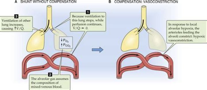
Improvement of the shunt fraction can be accomplished by decreasing blood flow or supplying O2 to the nondependent lung. Hypoxic pulmonary vasoconstriction is a powerful reflex that increases the PVR of the hypoxic lung and the atelectatic lung, diverting blood to the well-oxygenated areas of lung.
What does shunt mean in medical terms?(shunt) In medicine, a passage that is made to allow blood or other fluid to move from one part of the body to another. For example, a surgeon may implant a tube to drain cerebrospinal fluid from the brain to the abdomen.
Article first time published onIs anatomical shunt normal?
Anatomic shunt exists in normal lungs because of the bronchial and thebesian circulations, which account for 2-3% of shunt. A normal right-to-left shunt may occur from atrial septal defect, ventricular septal defect, patent ductus arteriosus, or arteriovenous malformation in the lung.
Is atelectasis a shunt?
The most common example of shunt is atelectasis, which is collapse of alveoli.
Is ARDS shunt or dead space?
Acute respiratory distress syndrome (ARDS) is characterized by severe impairment of gas exchange. Hypoxemia is mainly due to intrapulmonary shunt, whereas increased alveolar dead space explains the alteration of CO2 clearance.
Why Does dead space correct with oxygen?
Dead space is the volume of air that is inhaled that does not take part in the gas exchange, because it either remains in the conducting airways or reaches alveoli that are not perfused or poorly perfused. It means that not all the air in each breath is available for the exchange of oxygen and carbon dioxide.
How can toxic inhalation cause pulmonary edema?
If a blood clot travels from the blood vessels in your legs to your lungs, you can develop pulmonary edema. Exposure to certain toxins. Inhaling toxins or breathing in some of your stomach contents when you vomit (aspiration) causes intense irritation of the small airways and alveoli, resulting in fluid buildup.
Does shunt cause hypercapnia?
Shunt as a cause of hypoxemia is observed primarily in pneumonia, atelectasis, and severe pulmonary edema of either cardiac or noncardiac origin. Hypercapnia generally does not develop unless the shunt is excessive (> 60%).
What is a pulmonary shunt study?
A shunt study is a procedure to assess heart and lung blood circulation.
Which medication helps to treat infection in a patient with acute respiratory failure?
Antibiotics to treat infection. Anti-inflammatory drugs, such as corticosteroids, to reduce inflammation in the lungs in the late phase of ARDS or sometimes if the person is in septic shock. Diuretics to eliminate fluid from the lungs.
What is the difference between V Q mismatch and shunt?
A , VQ mismatch occurs with regional differences in the optimal alveolar-capillary interface as gas exchange occurs unimpeded (wide arrow) in some areas and restricted (narrow arrow) or prohibited (X) in others. … B , Shunt occurs when blood fl ow does not participate in gas exchange, such as is observed with ARDS.
What does a positive bubble test mean?
Bubble Test Results No bubbles should be seen on the far side of the heart. However, if bubbles do appear on the left side of the heart, this is a positive test and strongly indicates the presence of a hole in the heart.
When the lungs expand what happens to the intrapulmonary pressure?
Due to the adhesive force of the pleural fluid, the expansion of the thoracic cavity forces the lungs to stretch and expand as well. This increase in volume leads to a decrease in intra-alveolar pressure, creating a pressure lower than atmospheric pressure.
What are the five physiological causes of hypoxemia?
Hypoxemia is caused by five categories of etiologies: hypoventilation, ventilation/perfusion mismatch, right-to-left shunt, diffusion impairment, and low PO2.
What is shunt ventilation?
Shunt is defined as the persistence of hypoxemia despite 100% oxygen inhalation. The deoxygenated blood (mixed venous blood) bypasses the ventilated alveoli and mixes with oxygenated blood that has flowed through the ventilated alveoli, consequently leading to a reduction in arterial blood content.
How do you know if you have a hole in your heart?
- Shortness of breath.
- Easy tiring, especially after activity.
- Swelling of legs, feet or abdomen.
- Heart palpitations or skipped beats.
Is a right-to-left shunt bad?
A right-to-left shunt results in decreased blood flow through the pulmonary system, leading to decreased blood oxygen levels (hypoxemia). Hypoxemia manifests as cyanosis, causing “blue babies.”
What is a shunt in the brain?
A shunt is a hollow tube surgically placed in the brain (or occasionally in the spine) to help drain cerebrospinal fluid and redirect it to another location in the body where it can be reabsorbed.
Is a shunt in brain permanent?
Your doctor inserts one end of the tube in your brain and the other end into your chest or abdominal cavity. Excess fluid then drains from the brain and out the other end of the tube, where it can be more easily absorbed. A shunt implant is typically permanent and has to be monitored regularly.
What are the side effects of a brain shunt?
- redness and tenderness along the line of the shunt.
- a high temperature.
- headache.
- vomiting.
- neck stiffness.
- tummy pain if the shunt drains into your tummy.
- irritability or sleepiness in babies.
How often should a brain shunt be checked?
All younger patients with a shunt should probably be encouraged to seek a neurosurgical check up at least every three years, ideally at a dedicated hydrocephalus follow up clinic.
Is a PE a shunt?
Because pulmonary embolism (PE) alters perfusion rather than ventilation, it does not create an intrapulmonary shunt. By occluding the pulmonary vascular bed, however, PE can increase pulmonary vascular resistance and RV afterload, which can precipitate acute RV failure.
What causes hypercapnia?
Hypercapnia, or hypercarbia, is a condition that arises from having too much carbon dioxide in the blood. It is often caused by hypoventilation or disordered breathing where not enough oxygen enters the lungs and not enough carbon dioxide is emitted.