WHAT IS A Salter 1 fracture
A Salter-Harris type I fracture refers to a fracture line that runs straight across the growth plate, involving the cartilage without affecting the bone. Type I may cause the epiphysis, or the rounded end of the bone, to separate from the rest of the bone.
How long does it take for a Salter Harris type 1 fracture to heal?
Healing usually takes about 4-6 weeks, at which time it will be safe for your child to return to sports and activities. It is very rare for a Salter-Harris I fracture to cause problems with the growth of the distal fibula (less than 1% of fractures).
What are the 5 types of Salter Harris fractures?
- Salter I (Slipped) This is when the fracture line extends through the physis or within the growth plate. …
- Salter II (Above) These are when the fracture extends through both the physis and metaphysis. …
- Salter III (Lower) …
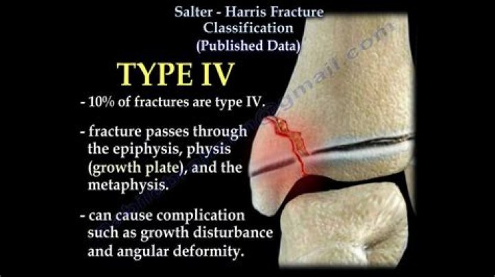
- Salter IV (Through/Transverse) …
- Salter V (Rammed/Ruined)
What is a Salter fracture?
A Salter-Harris fracture is a fracture in the growth plate of a child’s bone. A growth plate is a layer of growing tissue close to the ends of a child’s bone. It’s very important to get this condition diagnosed since it can affect a child’s growth.What is a Type 1 or 2 fracture?
They categorized open injuries into the familiar three categories, based on wound size, level of contamination, and osseous injury, as follows: Type I = an open fracture with a wound less than 1 cm long and clean; Type II = an open fracture with a laceration greater than 1 cm long without extensive soft tissue damage, …
Where does a boxer's fracture occur?
A boxer’s fracture is a break in the neck of the 5th metacarpal bone in the hand. It usually happens when you punch an object at a high speed. Symptoms of a boxer’s fracture include pain and swelling of the hand, limited range of motion of the pinky finger, and misalignment of the finger.
Does a Salter-Harris fracture need a cast?
Your child’s injury may need to be put in a cast or splint if a Salter-Harris fracture is known or suspected. This will help prevent more injury to the growth plate and surrounding bone. If the bone is not displaced (moved out of place), your child may get a cast to secure the bone as it heals.
What does Salter stand for?
S: slipped (type I) A: above or away from joint (type II) L: lower (type III) T: through or transverse or together (type IV) R: ruined or rammed (type V)What is a Salter-Harris Type 1 Physeal fracture?
Type 1. This fracture occurs when a force hits the growth plate separating the rounded edge of the bone from the bone shaft. It’s more common in younger children. About 5 percent of Salter-Harris fractures are type 1.
How many Salter fractures are there?There are nine types of Salter–Harris fractures; types I to V as described by Robert B Salter and W Robert Harris in 1963, and the rarer types VI to IX which have been added subsequently: Type I – transverse fracture through the growth plate (also referred to as the “physis”): 6% incidence.
Article first time published onWhere is Metaphysis located?
The metaphyses (singular: metaphysis) are the wide portions of long bones and the regions of the bone where growth occurs. Growth occurs at the section of the metaphysis that is adjacent to the growth plate (physis). The metaphysis is located between the diaphysis and epiphysis.
How do you treat a boxer's fracture?
The primary goal for medical treatment of a boxer’s fracture is to immobilize the hand to allow the bones to heal properly. Doctors usually employ various splints to do this job, though casts may be necessary, as well. The splint or cast should completely immobilize the joints above and below the site of the injury.
What is a Type II fracture?
A type II odontoid fracture is a break that occurs through a specific part of C2, the second bone in the neck. Bones of the spine are called vertebrae. The bone involved in odontoid fracture is the second vertebra, C2, high up in the neck.
What bones are included in a Lefort 1 fracture?
Bones fractured in a Le Fort I fracture include the lower nasal septum, the inferior portion of the pyriform apertures, the canine fossae, both zygomaticomaxillary buttresses, the posterior maxillary walls, and the pterygoid plates.
What are the four types of fractures?
- Stable fracture. The broken ends of the bone line up and are barely out of place.
- Open (compound) fracture. The skin may be pierced by the bone or by a blow that breaks the skin at the time of the fracture. …
- Transverse fracture. …
- Oblique fracture. …
- Comminuted fracture.
What are the 3 classifications of fractures?
- Open Fracture. When a broken bone breaks through the skin, it is classified as an open fracture. …
- Closed Fracture. …
- Displaced Fracture. …
- Subcategories.
What is metaphysis of bone?
The central tubular region of the bone, called the diaphysis, flares outward near the end to form the metaphysis, which contains a largely cancellous, or spongy, interior. … At the end of the bone is the epiphysis, which in young people is separated from the metaphysis by the physis, or growth plate.
Do spiral fractures require surgery?
Most spiral fractures require surgery and general anesthesia. Less severe cases, where the bone is not fully separated, may be operated on using local anesthesia. If the two ends of the bone are separated then an open reduction surgery will be necessary.
Which examination may be used to demonstrate a Salter-Harris fracture?
Taggart et al reported that the use of point-of-care ultrasonography in the emergency department setting could correctly diagnose Salter-Harris fractures. Findings of periosteal fluid at the level of the metaphysis and widening of the physis allowed for the diagnosis of a fracture.
Does a boxer's fracture hurt?
The typical symptoms of a boxer’s fracture are pain or tenderness on the hand near one of the metacarpal bones, around the knuckle. You may also have pain when you move your hand or fingers. When a bone is broken, you may have snapping or popping sensations.
Can you still box after a boxer's fracture?
Even when the strapping is removed the bone will not be strong enough for heavy work or non-contact sports until at least six weeks. Contact sports, in particular boxing, should be avoided until at least 12 weeks after injury. Sometimes these estimated times will take longer if pain and poor movement continues.
How long does a boxer's fracture hurt?
Ache – the hand often aches for 8–12 weeks after the fracture even though it has healed. In general it takes 6 weeks for a hand fracture to heal and a further 6 weeks to reach near normal strength Very heavy lifting and contact sport should be avoided until the fracture has solidly healed ( 8-12 weeks).
What are the complication of fracture?
Major complications of fracture repair include osteomyelitis, delayed union, nonunion, malunion, premature physeal closure, and fracture associated sarcoma. Consideration of these complications should factor into presurgical patient evaluation as well as postoperative management.
Is a closed reduction considered surgery?
Closed reduction is a procedure to set (reduce) a broken bone without surgery. It allows the bone to grow back together.
What is necessity fracture?
In 1941, Campbell termed the Galeazzi fracture the “fracture of necessity,” because it necessitates surgical treatment; in adults, nonsurgical treatment of the injury results in persistent or recurrent dislocations of the distal ulna.
What is the difference between Smith and Colles fracture?
Smith fractures do not extend to the wrist, hence they are extra-articular. These fractures are usually transverse. Smith fractures are rare and are most often seen in elderly women or young men. A Colles fracture is a broken wrist.
What is a buckle fracture in foot?
Buckle fractures are compression fractures and are very common in children. They happen when one side of the bone buckles, or bends, but doesn’t break all the way through. It is a stable fracture, meaning that the broken pieces of bone have not separated from each other.
When do your growth plates close?
Growth plates usually close near the end of puberty. For girls, this usually is when they’re 13–15; for boys, it’s when they’re 15–17.
Where is a growth plate located?
Growth plates, also called physes or epiphyseal plates, are discs of cartilage present in growing children. They are located between the middle and the end of the long bones, such as the bones of the arms and legs. Most long bones have one growth plate at each end.
What is cortical buckling?
Buckle (torus) fractures occur when the bony cortex is compressed and bulges, without extension of the fracture into the cortex (Figure 1). This type of fracture occurs in about 1 in 25 children and represents 50% of pediatric fractures of the wrist.
Where are the osteocytes?
Between the rings of matrix, the bone cells (osteocytes) are located in spaces called lacunae. Small channels (canaliculi) radiate from the lacunae to the osteonic (haversian) canal to provide passageways through the hard matrix.