What is mental protuberance
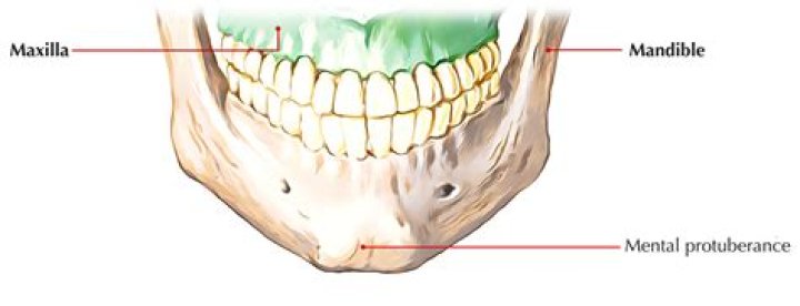
: the bony protuberance at the front of the lower jaw forming the chin.
What are mental tubercles?
Medical Definition of mental tubercle : a prominence on each side of the mental protuberance of the mandible.
Where are the mental tubercles located?
The symphysis of the external surface of the mandible divides below and encloses a triangular eminence, the mental protuberance, the base of which is depressed in the center but raised on either side to form the mental tubercle.
Is the mental protuberance a bone?
On the anterior inferior midline region of the hemimandible body is a triangular thickening of bone termed the mental protuberance. The thickened inferior rim of the mental protuberance extends laterally from the midline and forms 2 rounded protrusions termed the mental tubercles.What is the mental symphysis?
n. The fibrocartilaginous union of the two halves of an infant’s lower jawbone that ossifies during the first year.
Where is the Digastric fossa?
The digastric fossa of the mandible is an anatomical region which occupies a space on the inner surface of the inferior border of the body of the mandible, near the midline bilaterally. It is a position of attachment of the anterior belly of the digastric muscle.
What goes through mental foramen?
The mental foramen is located on the anterior surface of the mandible. It is directly below the commisure of the lips, and the tendon of depressor labii inferioris muscle. … It transmits the terminal branches of the inferior alveolar nerve (the mental nerve), the mental artery, and the mental vein.
What is the Forum Magnum?
The foramen magnum (Latin: great hole) is a large, oval-shaped opening in the occipital bone of the skull. It is one of the several oval or circular openings (foramina) in the base of the skull. … It also transmits the accessory nerve into the skull. The foramen magnum is a very important feature in bipedal mammals.What is the weakest part of the skull?
Clinical significance The pterion is known as the weakest part of the skull. The anterior division of the middle meningeal artery runs underneath the pterion. Consequently, a traumatic blow to the pterion may rupture the middle meningeal artery causing an epidural haematoma.
What is Pterygoid fovea?The pterygoid fovea is a small depression on the anteromedial surface of the condylar process of the mandible marking the attachment of the inferior belly of the lateral pterygoid muscle.
Article first time published onAre genial tubercles radiolucent or radiopaque?
Genial tubercle – a round/oval radiopaque structure inferior to the mandibular incisors. Hard palate – a radiopaque bony structure that separates the nasal cavity from the oral cavity.
Where is the condyle?
A condyle (/ˈkɒndəl/ or /ˈkɒndaɪl/; Latin: condylus, from Greek: kondylos; κόνδυλος knuckle) is the round prominence at the end of a bone, most often part of a joint – an articulation with another bone. It is one of the markings or features of bones, and can refer to: On the femur, in the knee joint: Medial condyle.
What is the tubercle of a bone?
A tubercle is a small rounded point of a bone. It also refers to a nodule attached to bone, mucous membrane (moist layer lining parts of the body), or skin.
What is unfused mandibular symphysis?
The mandibular symphyseal joint is remarkably variable across major mammalian clades, ranging in adults from unfused (amphiarthrosis) to partially fused (synarthrosis) to completely ossified (synostosis).
What species has a mandibular symphysis?
An ossified or ‘fused’ mandibular symphysis characterizes the origins of the Anthropoidea, a primate suborder that includes humans. Longstanding debate about the adaptive significance of variation in this jaw joint centers on whether a bony symphysis is stronger than an unfused one spanned by cartilage and ligaments.
What unites right and left halves of the mandible?
The right and left medial pterygoid plates form the posterior, lateral walls of the nasal cavity. The somewhat larger lateral pterygoid plates serve as attachment sites for chewing muscles that fill the infratemporal space and act on the mandible.
Why is the mental foramen important?
Background: The mental foramen is a strategically important landmark during osteotomy procedures. Its location and the possibility that an anterior loop of the mental nerve may be present mesial to the mental foramen needs to be considered before implant surgery to avoid mental nerve injury.
Can you palpate the mental foramen?
The mental foramen is situated on the lateral aspect of the ramus in the middle of the interdental space. It can be palpated after deflecting the pencil-like tendon of the depressor labii inferioris muscle upwards.
What is Ramus of mandible?
The rami are two vertical processes located on either side of the body; they join the body at the angle of the mandible. At the superior aspect of each ramus, the coronoid and condylar processes articulate with the temporal bone to create the temporomandibular joint which permits mobility.
What muscle is separated by two bellies?
Structure. The digastricus (digastric muscle) consists of two muscular bellies united by an intermediate rounded tendon. The two bellies of the digastric muscle have different embryological origins, and are supplied by different cranial nerves. Each person has a right and left digastric muscle.
What Innervates the Geniohyoid?
Innervation. The geniohyoid muscles are innervated by a branch of the ventral ramus of C1 from the cervical plexus which courses with the hypoglossal nerve in to the floor of the mouth.
What does the stylohyoid muscle do?
The stylohyoid muscle connects the hyoid bone to the base of the mandible and the skull. It pulls the hyoid bone upward and backward, resulting in elevation of the base of the tongue and elongation of the floor of the mouth. This movement helps in deglutition.
What is a Inion?
Definition of inion : the external occipital protuberance of the skull.
What lies deep Pterion?
The pterion is known as ‘the danger area’ on the skull for head injuries. This is because the bone is thin at this site and is grooved by vessels on its internal surface (or may even lie in a bony tunnel here). It is the anterior branch of the middle meningeal artery (and vein) that lies deep to the pterion.
Which bone is most superior?
The ilium is the largest and the most superior of the three bones that form the coxa (hipbone or hip joint).
What is the hypoglossal canal?
The hypoglossal canal is located between the occipital condyle and jugular tubercle and runs obliquely forwards (posteromedial to anterolateral) allowing the hypoglossal nerve to exit the posterior cranial fossa.
What is the only movable joint found in the skull?
The only bone in your skull that forms freely movable joints is your mandible, or jawbone.
Does the medulla pass through the foramen magnum?
occipital. occipital,, bone forming the back and back part of the base of the cranium, the part of the skull that encloses the brain. It has a large oval opening, the foramen magnum, through which the medulla oblongata passes, linking the spinal cord and brain.
What is mastication muscle?
The muscles of mastication are a group of muscles responsible for the chewing movement of the mandible at the temporomandibular (TMJ) joint, they enhance the process of eating, they assist in grinding food, and also function to approximate the teeth.
What is the Infratemporal fossa?
The infratemporal fossa is a space below the temporal fossa. Its borders include: Superiorly, the greater wing of the sphenoid bone. Inferiorly, the medial pterygoid muscle. Anteriorly, the maxilla.
What is the TMJ made of?
The main components are the joint capsule, articular disc, mandibular condyles, articular surface of the temporal bone, temporomandibular ligament, stylomandibular ligament, sphenomandibular ligament, and lateral pterygoid muscle.