What is the zygomatic bone
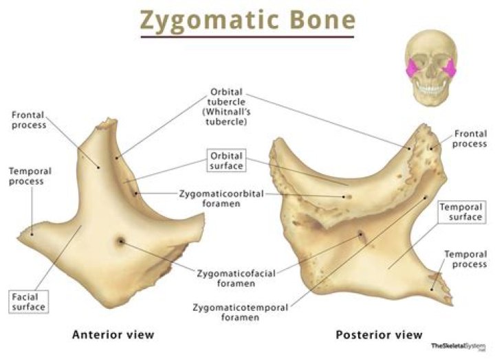
The zygomatic bones are a pair of diamond-shaped, irregularly-shaped bones that protrude laterally and form the prominence of the cheeks, a portion of the lateral wall, the orbit floor, and some portions of the temporal fossa and infratemporal fossa.
What part of the skeleton is the zygomatic bone?
The zygomatic bone (zygoma) is an irregularly shaped bone of the skull. It is often referred to as the cheekbone, and it comprises the prominence just below the lateral side of the orbit.
What is the zygomatic bone used for?
The zygomatic bone is a paired facial bone. Both zygoma or cheek bones are irregular and articulate with other bones of the cranium and face. They are important contributors to mastication or chewing, providing an attachment point for the masseter muscle – a jaw adductor that closes the jaw.
Where are the zygomatic and maxillary bones located?
In the human skull, the zygomatic bone (cheekbone or malar bone) is a paired irregular bone which articulates with the maxilla, the temporal bone, the sphenoid bone and the frontal bone.Where is a cheekbone?
One of a pair of bones on each upper side of the face that forms the cheek and part of the eye socket. The cheekbones help give shape and structure to the face and are connected to the jaw and bones near the ears, forehead, and skull.
What bones articulate with the zygomatic bones?
The zygomatic bone articulates with the sphenoid bone, maxilla, frontal bone, and temporal bone to form the lateral wall of the floor of the orbit, part of the temporal and infratemporal fossa, and the prominence of the cheek.
Why is it called zygomatic bone?
Zygomatic bone: The part of the temporal bone of the skull that forms the prominence of the cheek. … The word “zygomatic” comes from the Greek “zygon” meaning a yoke or crossbar by which two draft animals such as oxen could be hitched to a plow or wagon.
What is maxilla bone?
The maxilla is the bone that forms your upper jaw. The right and left halves of the maxilla are irregularly shaped bones that fuse together in the middle of the skull, below the nose, in an area known as the intermaxillary suture.What two bones make up the cheekbones?
- The zygomatic bones (Gr., zygoma – yoke) are two facial bones that form the cheeks and the lateral walls of the orbits.
- They are also commonly referred to a as the cheekbones or malar bones (L., mala – the cheek).
The temporal bones are two major bones in the skull, or cranium. They help form the sides and base of the skull, where they protect the temporal lobe of the brain and surround the ear canal. The other major bones in the skull are: the two parietal bones that make up the top of the skull.
Article first time published onWhere is sphenoid bone?
An unpaired bone located in the cranium (or skull), the sphenoid bone, also known as the “wasp bone,” is located in the middle and toward the front of the skull, just in front of the occipital bone.
What muscles originate from the zygomatic bone?
Origin(Posterior part of) Lateral aspect of zygomatic boneFunctionElevates and everts angle of mouth
What is zygomatic surgery?
Zygomatic also known as ‘Zygoma’ provide an alternative to bone grafting procedures in the upper jaw and are dental implants that transverse inside the poor area of your upper jaw to anchor into the underside of the cheekbones through your sinuses.
Where is the hyoid bone located?
The hyoid bone (hyoid) is a small U-shaped (horseshoe-shaped) solitary bone, situated in the midline of the neck anteriorly at the base of the mandible and posteriorly at the fourth cervical vertebra. Its anatomical position is just superior to the thyroid cartilage.
Does the zygomatic bone have a sinus?
It contains the largest of the paranasal sinuses, the maxillary sinus. To see the posterior part of the maxilla, we’ll remove the zygomatic arch. …
What bone forms the bridge of the nose?
Each of the following facial bones are paired: the maxillae form the upper jaw and front of the hard palate; the zygomatic bones form the cheeks; the nasal bones form the bridge of the nose; the lacrimal bones form part of the orbit, or eye socket; the palatine bones form the rear of the hard palate and the inferior …
What is the upper jaw bone called?
The upper part is the maxilla. It doesn’t move. The moveable lower part is called the mandible.
What 2 bones make up the zygomatic arch?
The facial area includes the zygomatic, or malar, bones (cheekbones), which join with the temporal and maxillary bones to form the zygomatic arch below the eye socket; the palatine bone; and the maxillary, or upper jaw, bones.
What happens if the zygomatic bone is damaged?
Fractures of the ZMC or zygomatic arch can often lead to unsightly malar depression, which should be corrected to restore a normal facial contour. ZMC fractures can also cause significant functional issues, including trismus, enophthalmos and/or diplopia, and paresthesias of the infraorbital nerve.
What causes pain in zygomatic bone?
Zygomatic arch pain is commonly reported by patients visiting the orofacial pain clinic and is majorly accepted to be caused by masseter muscle pain. But a variety of conditions may present as orofacial pain in the zygomatic arch region, including life-threatening diseases such as salivary gland tumors.
Is the zygomatic bone a flat bone?
Irregular Bones. They consist of cancellous tissue enclosed within a thin layer of compact bone. The irregular bones are: the vertebræ, sacrum, coccyx, temporal, sphenoid, ethmoid, zygomatic, maxilla, mandible, palatine, inferior nasal concha, and hyoid.
What are the names of the 22 bones in the skull?
- Frontal bone.
- Two parietal bones.
- Two temporal bones.
- Occipital bone.
- Ethmoid bone.
- Sphenoid bone.
What is the only movable bone in the skull?
The only bone in your skull that forms freely movable joints is your mandible, or jawbone.
What bone is the back of the skull?
The occipital bone is a bone that covers the back of your head; an area called the occiput. The occipital bone is the only bone in your head that connects with your cervical spine (neck).
What is the bone above your lip called?
MaxillaTA98A02.1.12.001TA2756FMA9711Anatomical terms of bone
What bones hold your teeth?
The alveolar bone is located on the jaw bones which hold the teeth. In humans, these bones that contain the teeth are the maxilla and the mandible. The curved portion of each alveolar process on the jaw is the alveolar arch.
Which bones are found on the temporal bone?
Temporal boneArticulationsOccipital, parietal, sphenoid, mandible and zygomaticIdentifiersLatinOs temporaleMeSHD013701
What is occipital bone?
The occipital bone is the most posterior cranial bone and the main bone of the occiput. It is considered a flat bone, like all other cranial bones, meaning that its primary function is either for protection or to provide a broad surface for muscle attachment. The scalp, which consists of five layers, covers the bone.
What is the location of the ethmoid bone?
The ethmoid bone is a cube-shaped bone located in the center of the skull between the eyes. It helps form the walls of the eye socket, or orbital cavity, as well as the roof, sides, and interior of the nasal cavity.
Where is sella turcica located?
The sella turcica is a saddle-shaped depression located in the bone at the base of skull (sphenoid bone), in which resides the pituitary gland.
Where is the foramen ovale located in the skull?
The foramen ovale is an oval shaped opening, placed obliquely in the base of the skull. It is situated in the greater wing of sphenoid bone, close to the upper end of posterior margin of lateral pterygoid plate, medial to foramen spinosum and lateral to the foramen lacerum [1].